Nuclear Medicine is a medical speciality that uses safe, painless, and cost-effective
techniques both to image the body and treat disease.
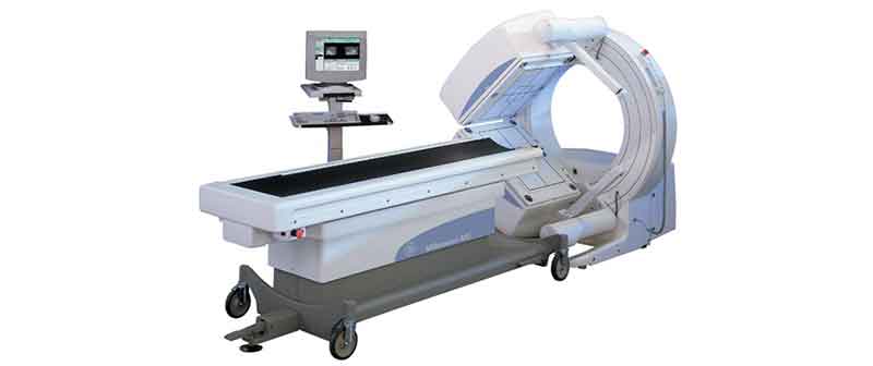
Nuclear imaging is unique in that it documents both organ function and morphology.Gamma ray emitting radiotracer is injected intravenously and non-invasive imaging is performed with state-of-the-art Dual Head Gamma Camera (GE Millennium VG)
When we already have X-ray, Ultrasound, CT & MRI, why do we need this?
Radionuclide procedures are available for almost all organ systems just like the different X-ray procedures. It is used in evaluation of Heart, Lung, Brain, Bone, Kidneys, Liver etc.Scans are designed to study various aspects like concentration, excretion, drainage/flow of tracers in various organs or localize/characterize the leison pathology.
The value of Whole body Positron Emission Tomography–Computed Tomography (PET/CT) for diagnostic imaging in oncology is well established with its capabilities to combine anatomical and functional aspects of the whole body to provide a complete picture of patient’s disease status.
Most common uses of PET/CT are found in Oncology, Cardiology and Neurology practices.
Millennium MG, Multi-Geometry Nuclear Medicine System is used to perform a range of Nuclear Imaging studies. Imaging with Gamma Camera involves preparation and injection of radiopharmaceuticals that specifically trace the function of Organ of Interest. Radiopharmaceuticals are prepared in the well-equipped Hot Lab in the Department of Molecular Imaging. These specific radiopharmaceuticals are administered to patients and images are acquired. Static, dynamic and parametric images can be obtained under a Gamma Camera.
Myocardial Perfusion Scan visualizes the distribution of tracer uptake in the heart muscle, which reflects regional blood flow in different coronary artery territories. This can be performed with 201T1/ 99m Tc- Sestamibi / Trofosmin.
Rest Injection Scan indicates whether the heart muscle is viable or scarred due to prior attacks. Stress (Exercise/Pharmacological) Scan reveals inducible ischemic perfusion defect corresponding to significant coronary disease.
A bone scan is a diagnostic imaging study which records the distribution of a radioactive tracer in the skeletal system. It is the most sensitive study available to pick up any pathology of the skeleton.
Renal scans are performed to evaluate the differential renal function, the extraction and excretion function of the kidneys. Information about Glomerular Filtration Rate (GFR), tubular function and cortical morphology can be obtained by such scans.
Brain Perfusion SPECT with advanced NeuroGam Analysis Package for diagnosing
Head & Consultant in Dept. of Nuclear Medicine and Molecular Imaging